Mitral valve prolapse (MVP) occurs during ventricular contraction (systole). Specifically, during systole, the increased pressure in the left ventricle causes the mitral valve leaflets to billow or prolapse back into the left atrium. This abnormal systolic displacement of the mitral leaflets beyond the annular plane leads to mitral regurgitation in many cases.
The echocardiographic hallmark of MVP is systolic bowing or displacement of the mitral leaflets into the left atrium, best visualized in parasternal long-axis or apical views during ventricular contraction. MVP is not seen during ventricular filling phases such as early filling, atrial systole, or diastasis because the leaflets are normally open or positioned differently.
This is well-documented in the "Textbook of Clinical Echocardiography, 6e", Chapter on Mitral Valve Disease, explaining the pathophysiology of MVP and its timing during the cardiac cycle【20:390-395†Textbook of Clinical Echocardiography】.
Question 41
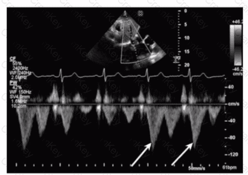
Which flow component is indicated by the arrows on this image?
The Doppler waveform shows pulmonary vein flow with several components. The arrows point to small reversed flow spikes just after the atrial contraction wave, which corresponds to the atrial reversal (AR) flow component. Atrial reversal occurs as blood briefly flows backward into the pulmonary veins during atrial contraction.
Ventricular reversal is not typically seen in pulmonary veins. Diastolic flow reversal is abnormal and usually not part of normal pulmonary vein flow. Systolic forward flow is the major forward component during ventricular systole.
This interpretation is standard in ASE guidelines on diastolic function assessment and pulmonary vein Doppler evaluation【12:ASE Diastolic Function Guidelines†p.85-90】【16:Textbook of Clinical Echocardiography, 6e†p.130-135】.