Yes, very much so. Cramkey Dumps are created by experienced and certified professionals who have gone through the exams themselves. They understand the importance of providing accurate and relevant information to help you succeed.
Mylo
Excellent dumps with authentic information… I passed my exam with brilliant score.
DominikMay 10, 2026
That's amazing! I've been looking for good study material that will help me prepare for my upcoming certification exam. Now, I will try it.
Miriam
Highly recommended Dumps. 100% authentic and reliable. Passed my exam with wonderful score.
MilanMay 26, 2026
I see. Thanks for the information. I'll definitely keep Cramkey in mind for my next exam.
Alaia
These Dumps are amazing! I used them to study for my recent exam and I passed with flying colors. The information in the dumps is so valid and up-to-date. Thanks a lot!!!
ZofiaMay 28, 2026
That's great to hear! I've been struggling to find good study material for my exam. I will ty it for sure.
Mariam
Do anyone think Cramkey questions can help improve exam scores?
KatieMay 10, 2026
Absolutely! Many people have reported improved scores after using Cramkey Dumps, and there are also success stories of people passing exams on the first try. I already passed this exam. I confirmed above questions were in exam.
Question 13
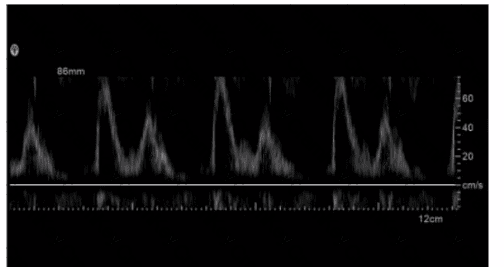
Which adjustment is needed to optimize the waveform below?
The waveform in the image shows spectral Doppler signals that are pushed against the upper limit of the display, indicating that the baseline is too high. Lowering the baseline allows for a better visual representation of the entire Doppler signal within the available display range. This adjustment prevents the waveform from being cut off and helps in accurately interpreting the blood flow characteristics.
Increased Transmit Frequency: This would generally improve the resolution of the image but does not directly correlate to the changes seen in the provided image link.
Increased Scale: Adjusting the scale changes the velocity range displayed but does not directly affect the speckle or noise reduction.
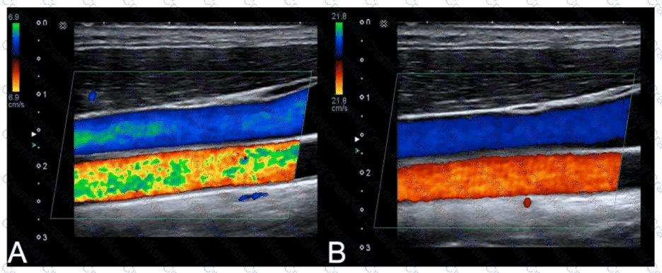
Decreased Color Gain: Reducing the color gain can decrease the amount of color noise, making the blood flow regions more defined, which aligns with the change observed from image A to image B.
Decreased Acoustic Power: This reduces the overall intensity of the ultrasound beam, affecting penetration depth and overall brightness but is less likely to result in the specific improvements seen.
[References:, "Understanding Ultrasound Physics" by Sidney K. Edelman, ARDMS Sonography Principles and Instrumentation study materials, , , ]
Question 15
Which artifact displays reflectors more shallow than their actual position?
Range ambiguity artifact occurs when echoes from one pulse are received after the next pulse has been emitted, leading to the incorrect placement of echoes at shallower depths than their true location. This artifact typically happens when the PRF is set too high, causing the ultrasound system to interpret delayed echoes as coming from the current pulse rather than the previous one. This results in reflectors appearing closer to the transducer than they actually are.